EOS
EOS : 3D Imaging device in standing and sitting position
EOS outstanding features
● Based on the Nobel prize winning detection technology by George Charpak in 1992
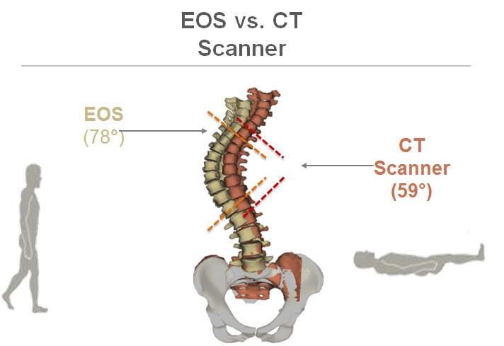
● acquiring images in weight bearing position
● Major reduction in radiation exposure compared to CT images (500 to 1000 times lower)
● Capable of producing 3D images out ofaccurate 2D images
● Enhanced Imaging speed and accuracy
● Producing life-size images
● Providing the physician with more than 100 important parameters about the natural and accurate size and angles of the skeleton
Unique Imaging technology
EOS imaging device includes two X-ray tube and two linear detectors which are placed in a cylindical room and move from bottom to top to take images from
frontal amnd lateral sides. The imaging can be executed locally or globally (specific parts of body or overal imaging). A whole body scan takes up to 15-20
seconds. The imaging can be done in standing, sitting and limped position.
Low dose imaging technology of EOS is based on the Nobel winning technology invented by chaprak. This images are taken by a method called Slot-Scanning that can reduce the amount of scattered rays and consequently increase the S/N ratio and dynamic range of the device. These two features help produce a high quality image with substantial lower dose.
SterEOS®, Surface 3D modelling software
EOS® 3D images are created via patented sterEOS® software from two unique low dose images, with no additional radiation. The algorithms are based on statistical modeling and bone shape recognition. This modeling falicitates the automatic calculation of many tridimensional clinical parameters. 3D skeletal envelope images can be obtained for the spine, the femur and the tibia. Unlike CT, EOS® images are acquired in an upright position, enabling new ways to globally evaluate a patient's postural abnormality.
SterEOS® 3D modeling allows the display of bone position, rotation and orientation. It also enables the display in different perspectives, in conjunction with the calculation of over 100 clinical parameters relevant for surgical planning and follow-up of pathologies..
Clinical parameters calculated automatically with sterEOS® include Cobb angle, vertebral axial rotation, saggital balance parameters, pelvic parameters, neck shaft angle, 3D femoral-tibial angles and lengths, rotational and torsional parameters of the lower limb.
EOS applications:
● Analysis of spine deformities like Deforming dorsopathies, Scoliosis, Lordosis, Kyphosis
● Diagnosis of knee, thigh and other lower limbs conditions
● Further analysis of patient skeletal condition before major surguries like total hip and total knee replacement
● Analysis of implants performance in patient body
● Analysis of torsion in lower lims
● Analysis of osteoporosis fractures
● Sagittal balance analysis
● When the patient should not be exposed to x-ray radiation
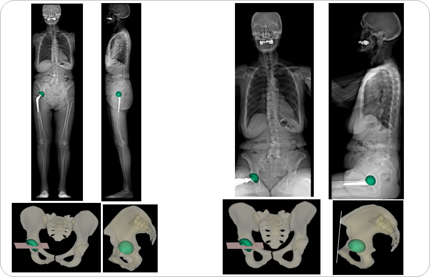
A few samples of the images taken by EOS imaging device